Interferometric image scanning microscopy (iISM) is a new label-free imaging method from W.E. Moerner’s lab at Stanford that can see structures inside living cells at about 120 nanometers of resolution without fluorescent dyes. It combines interferometric scattering microscopy, in which a laser makes tiny cell structures scatter light and a second beam amplifies that weak signal through interference, with the array detector used in advanced confocal microscopes, which collects tens to hundreds of slightly different views that software merges into one sharper image.
Because it needs no labels, iISM avoids photobleaching, can follow many cell components at once over long periods, and uses substantially less light than comparable label-free methods, which lowers the risk of photodamage. The researchers describe it as a complement to fluorescence microscopy rather than a replacement.
Source: “Stanford’s new microscope reveals living cells like never before,” ScienceDaily, from materials provided by Stanford University (October 16, 2026); study: Küppers and Moerner, “Interferometric Image Scanning Microscopy for label-free imaging at 120 nm lateral resolution inside live cells,” Light: Science & Applications 15(1) (2026), DOI 10.1038/s41377-026-02210-y.
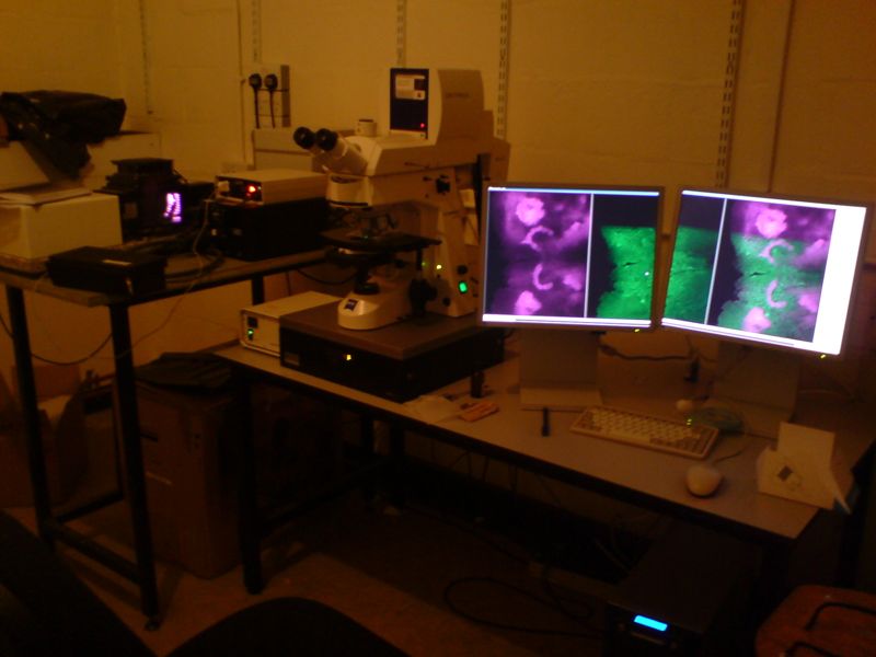
Image: A confocal microscope workstation (not the Stanford iISM instrument). Mark from Edinburgh, United Kingdom, CC BY-SA 2.0, via Wikimedia Commons.